Radiology is a field of medicine that uses imaging technology, like X-rays and other advanced machines, to diagnose cancer and other diseases. At Fox Chase Cancer Center, board-certified radiologists are a critical part of the medical team, participating in the diagnosis, staging and surveillance of all types of tumors.
Imaging Techniques Available at Fox Chase
Ultrasound
An ultrasound is a test that uses sound waves to create an image of internal organs. Abnormal tissue growth creates different sound-wave echoes than healthy tissue, allowing radiologists to locate potential tumors. Ultrasound is often used to guide biopsies.
CT Scan
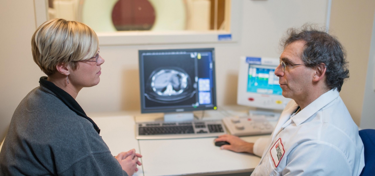
A computed tomography (CT) scan is a common imaging test used to detect and stage cancer by creating 2D and 3D pictures of the inside of the body. CT scans can reveal tumors and be used to evaluate if a treatment is working. They can also be used to guide biopsies and for placement of fiducial markers for radiation treatment.
MRI Scan
A magnetic resonance imaging (MRI) test uses magnets and radio waves to create detailed images of tissues inside the body. A radiologist may use an MRI to locate a tumor and determine its size and location—and whether it’s benign or malignant. Fox Chase specialists also use MRI to guide breast and prostate biopsies.
PET-CT and Nuclear Medicine Studies
Positron emission tomography with CT (PET-CT) is an imaging test that provides doctors with information about the body’s chemistry, cell function and exact location of disease.
Learn more about PET-CT and nuclear medicine studies →
X-ray/Fluoroscopy
An X-ray is an imaging test that uses radiation to create pictures of the inside of the body. A fluoroscopic study is a specialized kind of X-ray that creates a “movie” out of continuous X-ray images. Fluoroscopy at Fox Chase is often used to identify a problem with swallowing or with the stomach or bowel.
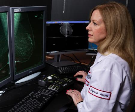
Breast Imaging
Fox Chase Cancer Center is designated as a Breast Imaging Center of Excellence by the American College of Radiology. Our breast imaging center offers mammography with 3D tomosynthesis, breast MRI, breast ultrasound, stereotactic biopsy, MRI and ultrasound-guided breast biopsy. We also perform localization of breast tumors before surgery.
Learn more about breast imaging →
Virtual Colonoscopy (CT Colonography)
A virtual colonoscopy is just what it sounds like–a noninvasive test that uses high-quality images and specialized software to “virtually examine” the colon without having to physically insert a scope into a patient.
Biopsies
In addition to imaging, Fox Chase radiologists specialize in performing challenging image-guided biopsies. Using ultrasound, CT or MRI guidance, radiologists can sample even small lesions or lesions that are difficult to access by other means, as well as biopsies on patients with complicating medical factors. A biopsy uses a small needle to remove tiny pieces of tissue from the lung, liver, lymph nodes or other body parts. Biopsies are used to diagnose or stage cancer, or for molecular testing to better understand the makeup of a tumor. All biopsies performed at Fox Chase are minimally invasive.