
Nuclear medicine is a type of imaging that uses very small amounts of radioactive material to diagnose and track diseases, including many types of cancer. These tests don’t simply take pictures; they can also tell doctors how an organ is functioning.
The most common areas examined during a nuclear medicine test include the bones, kidneys, lungs, thyroid and prostate. Sentinel node mapping is an important procedure to help surgeons localize lymph nodes for sampling, especially for breast cancer, melanoma, anal cancer and many gynecologic tumors.
During a nuclear medicine test, patients swallow, inhale or are injected with a radioactive substance (called a tracer). This tracer gathers in the organ, bone or tissue that doctors want to examine. Once there, the tracer helps “light up” those areas so that advanced cameras can take detailed pictures of them. These images help document the structure and function of the tissue or organs of interest. Nuclear medicine procedures are often longer than other imaging studies since the pictures are often taken continuously.
In some cases, nuclear medicine procedures are used as therapies, and as a Comprehensive Cancer Center, Fox Chase provides a wide spectrum of treatment options. The therapies include the use of radioactive iodine to treat hyperthyroidism or thyroid cancer. Other nuclear medicine therapies can help relieve bone pain with Xofigo for prostate cancer and Samarium for other cancers. We also provide Zevalin treatment for Non-Hodgkin’s Lymphoma, and liver directed therapy with Sirsphere and Therasphere.
Nuclear medicine tests are noninvasive and require little to no preparation on the patient’s part. Procedure times vary depending on the test.
PET-CT Scan
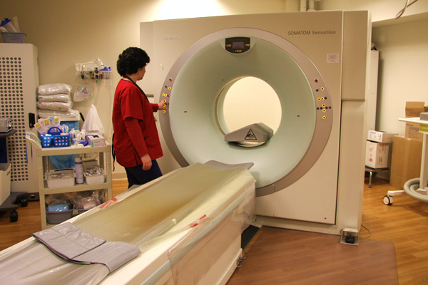
Positron emission tomography (commonly called a “PET scan”) is an advanced imaging machine used by nuclear medicine specialists to diagnose or follow certain cancers. A PET scan can often detect tumors before they show up on other imaging tests like CT or MRI.
The most advanced imaging technology today is PET and CT together in one integrated scanner. This combines the images from a PET scan and a computed tomography (CT) scan that have been performed at the same time using the same machine. Together, these two scans create a more accurate picture of what is going on in the body than either test can offer alone.
PET scans are often performed to:
- Detect cancer
- Help to point out the site for biopsy/tissue sampling
- Determine whether a cancer has spread in the body
- Assist with radiation planning and modification
- Assess the effectiveness of a treatment plan, such as cancer therapy
- Determine if a cancer has returned after treatment
- Participate in many research studies
Scanning for Recurrence in Prostate Cancer Patients
Fox Chase Cancer Center is the first facility in the region to offer a new clinical service for prostate cancer patients who are concerned about recurrence. The procedure uses a new imaging agent, fluorine-18-labeled synthetic amino acid for PET scans, which uses a radioactive drug to reveal how tissue and organs are functioning. In men with suspected prostate cancer recurrence due to an elevated PSA (prostate specific antigen) level, this drug uses the elevated levels of amino acids in cancer cells to help identify the location of a cancer recurrence. It is now available as a clinical service in the PET center.