This Fox Chase professor participates in the Undergraduate Summer Research Fellowship.
Learn more about Research Volunteering.

This Fox Chase professor participates in the Undergraduate Summer Research Fellowship.
Learn more about Research Volunteering.
Professor/Senior Member
Carol & Kenneth E. Weg Chair in Human Genetics
Chief, Genomic Medicine
Director, Clinical Cytogenomics Laboratory


Director, Genomics Facility
Molecular biology of mesothelioma; role of AKT in oncogenesis
The Testa lab focuses on two research areas: 1) hereditary and somatic genetics of malignant mesothelioma, a cancer of the serosal cells lining the chest and abdominal cavities, primarily caused by exposure to asbestos; and 2) the role of AKT oncogenes in tumorigenesis. The lab integrates data from human primary tumor tissues and tumor-derived cell lines as well as tumors from genetically engineered mouse models (GEMM) to address research questions in these two subject areas. As a result of the group’s interest in hereditary aspects of mesothelioma, the lab discovered the first germline (heritable) BAP1 mutations in two families with a high incidence of mesothelioma, and the group observed somatic alterations affecting BAP1 in familial mesotheliomas, indicating biallelic inactivation. In addition to mesothelioma, some BAP1 mutation carriers developed uveal melanoma or other cancers, consistent with the existence of a novel tumor syndrome, now known as BAP1 tumor predisposition syndrome (BAP1-TPDS). The group is also investigating other genes that may contribute to mesothelioma susceptibility and progression. GEMM are being used to assess gene–environment interactions in mesothelioma pathogenesis and as preclinical models for targeting cellular pathways important in this disease. With regard to AKT, the lab continues to study the role of the AKT1 and AKT2 oncogenes in tumorigenesis and drug resistance to therapies.
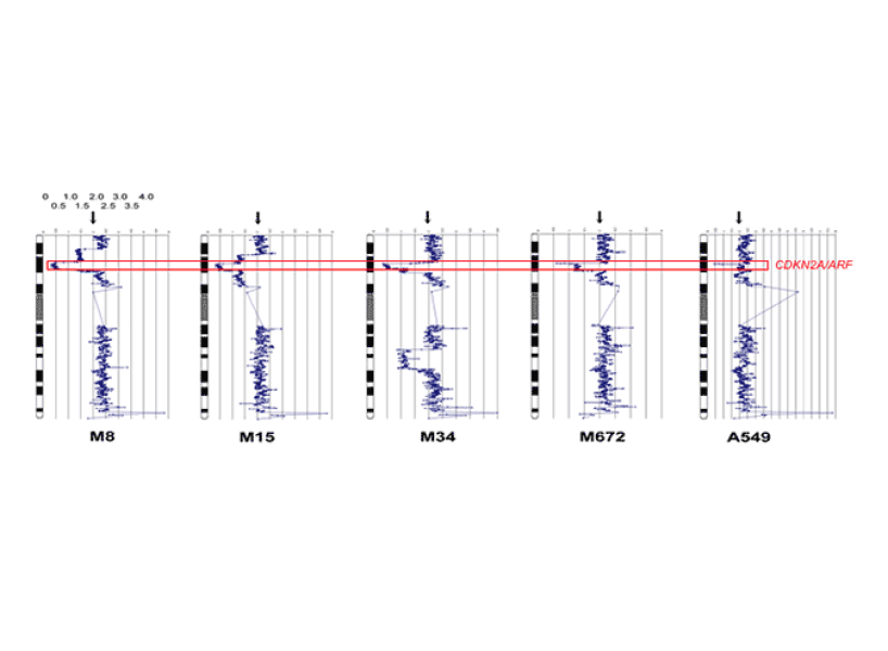
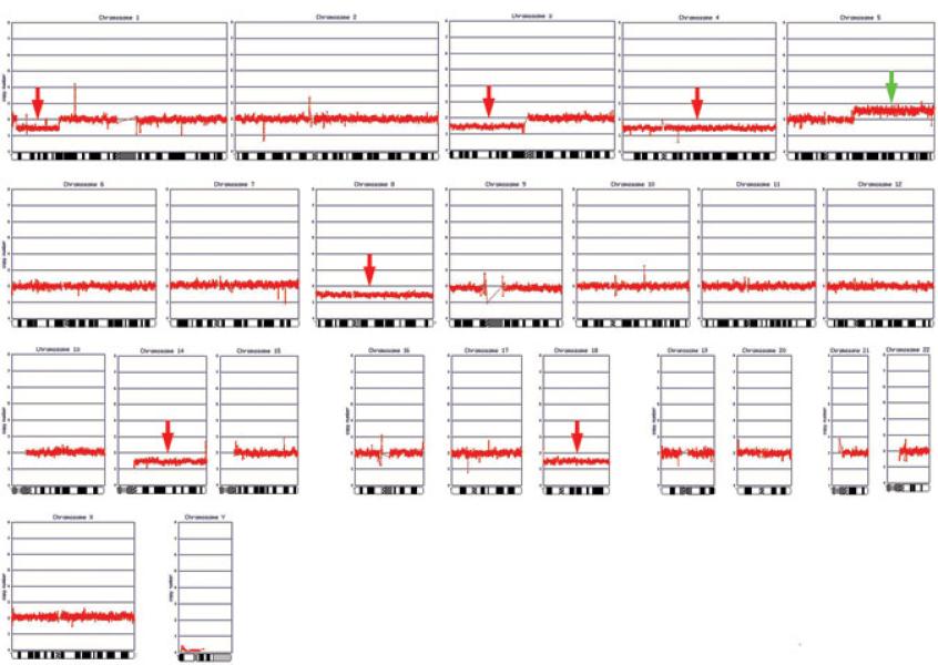
The Testa lab investigates the role of hereditary and somatic mutations in malignant mesothelioma, an incurable form of cancer often caused by exposure to asbestos. The group discovered frequent mutations of the CDKN2A locus, a region of DNA that encodes the tumor suppressors p16INK4A and p14ARF, and NF2/merlin in human mesothelioma. In 2011, he and his collaborators also discovered germline mutations of the BAP1 tumor suppressor gene in families with a high incidence of mesothelioma, the first study demonstrating that inherited mutations can influence a person’s risk of mesothelioma. Besides mesothelioma, some of the BAP1 mutation carriers developed ocular melanoma or other cancers, and this cancer susceptibility is now recognized as the BAP1 tumor predisposition syndrome (BAP1-TPDS). The Testa lab has reported multiple families with the BAP1-TPDS, including some family members with two or more different types of cancer, suggesting widespread BAP1-related tumor susceptibility targeting tissues of multiple organs. One of the families with a germline nonsense mutation in BAP1 included five relatives with peritoneal mesotheliomas as well as second or third primary cancers, including two with meningiomas. Two family members had basal cell carcinomas, and six others had melanocytic tumors, including four cutaneous melanomas, one uveal melanoma, and one benign melanocytic tumor. Given that this family resides in a subtropical area, and several members had suspected exposure to asbestos either occupationally or in the home, we hypothesized that the concurrence of a genetic predisposing factor and environmental exposure to asbestos and UV irradiation contributed to the high incidence of multiple cancers seen in this family, specifically mesothelioma and various uveal/skin tumors, respectively.
In another, large study, the Testa lab examined the germline BAP1 mutation status of 150 mesothelioma patients with a family history of cancer, 50 asbestos-exposed control individuals with a family history of cancers other than mesothelioma, and 153 asbestos-exposed individuals without familial cancer. No BAP1 alterations were found in control cohorts, but were identified in 9 of 150 mesothelioma cases (6%) with a family history of cancer. Alterations among these cases were characterized by both missense and frameshift mutations, and enzymatic activity of BAP1 missense mutants was decreased compared with wild-type BAP1. Furthermore, BAP1 mutation carriers developed mesothelioma at an earlier age that was more often peritoneal than pleural (5 of 9) and exhibited improved long-term survival compared to mesothelioma patients without BAP1 mutations. Moreover, many tumors harboring BAP1 germline mutations were associated with the BAP1-TPDS. Collectively, these findings suggest that mesothelioma patients presenting with a family history of cancer should be considered for BAP1 genetic testing to identify those individuals who might benefit from further screening and routine monitoring for the purpose of early detection and intervention. In a subsequent international intergroup study that included the Testa lab, 181 BAP1-TPDS families worldwide were reviewed, and the collated data confirmed the core tumor spectrum associated with the syndrome. Median ages of onset of core tumor types were lower in null than missense variant carriers for all tumors combined, mesothelioma, cutaneous melanoma, and nonmelanoma skin cancer (each having P values of < 0.001). In addition to BAP1, the Testa lab is also investigating other genes that may predispose to mesothelioma.
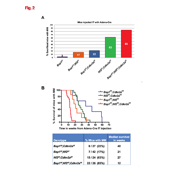
Much of the current work in the lab is focused understanding the mechanisms involved in BAP1-related tumor susceptibility. Using several GEMM they developed, the Testa lab provided the first in vivo evidence that heritable (germline) mutations of Bap1 predispose to the development of asbestos-induced mesothelioma. This and other Bap1-mutant mouse models are currently being used to assess susceptibility to spontaneous mesothelioma formation and gene–environment interactions. In one project, the Testa lab introduced various combinations of deletions of Bap1, Cdkn2a, and/or Nf2 in the pleura of conditional knockout (CKO) mice, focusing on the contribution of Bap1 loss. While homozygous CKO of Bap1, Cdkn2a, or Nf2 alone gave rise to few or no mesotheliomas, inactivation of Bap1 cooperated with loss of either Nf2 or Cdkn2a to drive development of mesothelioma in approximately 20% of double-CKO mice, and a high incidence (22/26, 85%) of mesotheliomas was observed in Bap1;Nf2;Cdkn2a (triple)-CKO mice. Malignant mesothelioma onset was rapid in triple-CKO mice, with a median survival of only 12 weeks, and the mesotheliomas from these mice were consistently high-grade and invasive. Adenoviral-Cre treatment of normal mesothelial cells from Bap1;Nf2;Cdkn2a CKO mice, but not from mice with knockout of one or any two of these genes, resulted in robust spheroid formation in vitro, suggesting that mesothelial cells from Bap1;Nf2;Cdkn2a mice have stem cell-like potential. RNA-seq analysis of mesotheliomas from triple-CKO mice revealed enrichment of genes transcriptionally regulated by the polycomb repressive complex 2 (PRC2) and others previously implicated in known Bap1-related cellular processes. These data demonstrate that somatic inactivation of Bap1, Nf2, and Cdkn2a results in rapid, aggressive mesotheliomas, and that deletion of Bap1 contributes to tumor development, in part, by loss of PRC2-mediated repression of tumorigenic target genes and by acquisition of stem cell potential, suggesting a potential avenue for therapeutic intervention.
The group is also using novel GEMM to unravel the role of asbestos-induced inflammation in the genesis of malignant mesothelioma. For example, the Testa lab demonstrated that inflammation-related IL1β/IL1R signaling promotes the onset of asbestos-induced malignant mesothelioma, and the team’s in vivo findings provided rationale for chemoprevention strategies targeting IL1β/IL1R signaling in high-risk, asbestos-exposed populations. Ongoing preclinical studies with mouse models are being performed to assess the efficacy of certain other anti-inflammatory drugs as chemoprevention agents.
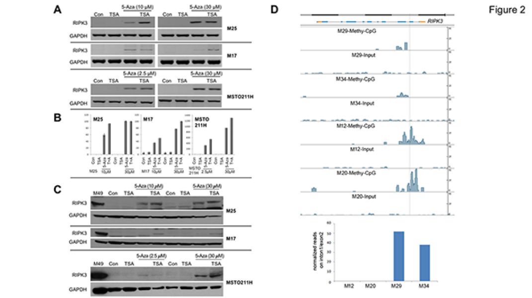
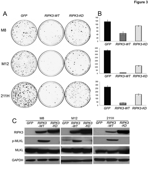
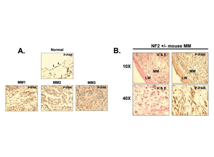
Mechanistic work by the Testa lab previously linked NF2 inactivation to oncogenic PAK and FAK signaling, implicating NF2 inactivation in mesothelioma cell spreading, invasiveness and proliferation, thereby establishing a framework for elucidating tumorigenic mechanisms and novel therapeutic targets in this disease. Other in vivo studies demonstrated that genetically engineered deletions of Nf2 and Cdkn2a cooperate to drive the development of highly aggressive mesotheliomas characterized by enhanced tumor dissemination and the involvement of a cancer stem cell (CSC) population. Mesothelioma is very difficult to treat and almost always recurs after therapy. Based on his work linking NF2 loss to FAK activation, he partnered with investigators in the pharmaceutical industry to test a novel drug that blocks FAK activity. Their findings suggested that FAK inhibitor treatment might be especially beneficial in patients with NF2-deficient tumors. Moreover, the drug proved to be particularly effective at killing CSCs, which can give rise to recurrent tumors. These preclinical studies provided the rationale for a clinical trial in mesothelioma patients using a FAK inhibitor as a single agent after first-line chemotherapy. More recently, other mechanistic studies have demonstrated that RIPK3, which encodes a receptor-interacting protein kinase, acts as a tumor suppressor in mesothelioma by triggering necroptosis, and that epigenetic silencing of RIPK3 by DNA methylation inactivates necroptosis and contributes to chemoresistance and poor survival in this incurable disease.
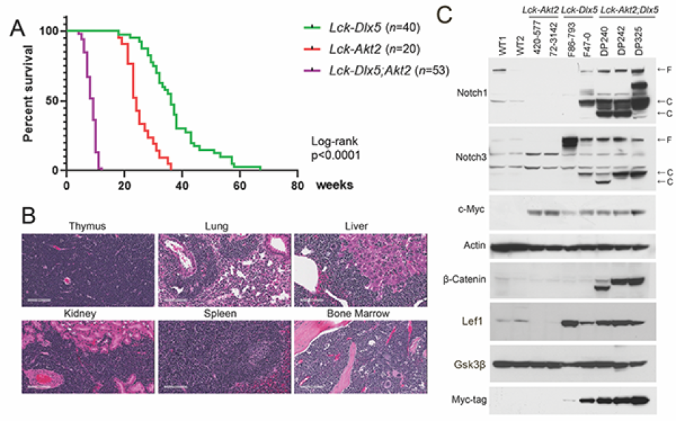
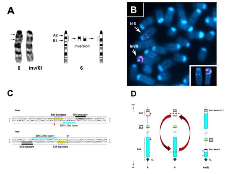
Testa has had a longstanding interest in the oncogenic role of AKT, beginning with his chromosomal mapping of the AKT1 proto-oncogene in 1988. The following year, Testa brought the AKT project to Fox Chase, when he joined the Center and jointly published a series of seminal studies with Phil Tsichlis and Alfonso Bellacosa. In a highly cited 1991 paper in Science, the team reported that the retroviral oncogene, akt, encodes a predicted oncoprotein contained viral Gag sequences fused to a serine-threonine kinase related to protein kinase C. The oncogenic potential of v-akt arises from the creation of a myristylation site at the amino terminus and consequent constitutive kinase activity, whereas the cellular homolog c-akt requires mitogenic stimulation for its activation. The Testa lab cloned and characterized the related AKT2 gene and provided the first evidence for recurrent alterations of the AKT pathway in human cancers. In recent years, the lab has characterized how other genes, such as the proto-oncogene Myc or the homeobox gene Dlx5 cooperate with Akt2 to promote T-cell lymphomagenesis. Recently, the Testa lab generated compound Dlx5;Akt2 transgenic mice, and the team discovered that activated Akt2 synergized with over-expressed Dlx5 to greatly accelerate and enhance the dissemination of T-lymphomagenesis. RNA-seq analysis performed on lymphomas from these mice revealed upregulation of genes involved in the Wnt and cholesterol biosynthesis pathways. Combined RNA-seq and ChIP-seq analysis of lymphomas from Dlx5;Akt2 mice demonstrated that β-catenin directly regulates genes involved in sterol regulatory element binding transcription factor 2 (Srebf2)-cholesterol synthesis. These lymphoma cells had high Lef1 levels and were highly sensitive to β-catenin and Srebf2-cholesterol synthesis inhibitors. Similarly, human T-cell leukemia/lymphoma (T-ALL) cell lines with activated NOTCH and AKT and elevated LEF1 levels were sensitive to inhibition of β-catenin and cholesterol pathways. Furthermore, LEF1 expression positively correlated with expression of genes involved in the cholesterol synthesis pathway in primary human T-ALL specimens. Together, these data suggest that targeting β-catenin and/or cholesterol biosynthesis, together with AKT, could have therapeutic efficacy in a subset of T-ALL patients.
University of Pennsylvania
Krais J., Glass D., Chudoba I., Wang Y., Feng W., Simpson D., Patel P., Liu Z., Neumann-Domer R., Betsch R., Bernhardy A., Bradbury A., Conger J., Yueh W.-T., Nacson J., Pomerantz R., Gupta G., Testa J.R., Johnson N. Genetic separation of Brca1 functions reveal pre-mitotic RPA as a driver of Polq addiction. Nat Commun. (in press).
Kadariya Y., Sementino E., Shrestha U., Gorman G., White J.M., Ross E.A., Clapper M.L., Neamati N., Miller M.S., Testa J.R. Inflammation as a chemoprevention target in asbestos-induced malignant mesothelioma. Carcinogenesis 43:1137-48, 2022. PMID: 36355620 PMCID: PMC10122428.
Osmanbeyoglu HU, Palmer D, Sagan A, Sementino E, Becich MJ, Testa JR. Isolated BAP1 genomic alteration in malignant pleural mesothelioma predicts distinct immunogenicity with implications for immunotherapeutic response. Cancers (Basel). 16;14:5626, 2022. PMID: 36428720; PMCID: PMC9688367.
Kurimchak A.M., Herrera-Montávez C., Montserrat-Sangrà S., Araiza-Olivera D., Hu J., Neumann-Domer R., Kuruvilla M., Bellacosa A., Testa J.R., Jin J., Duncan J.S. The drug efflux pump MDR1 promotes intrinsic and acquired resistance to PROTACs in cancer cells. Sci Signal 15:eabn2707, 2022. PMID: 36041010 PMCID: PMC9552188.
Sementino E., Kadariya Y., Cheung M., Menges C.W., Tan Y., Kukuyan A.M., Shrestha U., Karchugina S., Cai K.Q., Peri S., Duncan J.S., Chernoff J., Testa J.R. Inactivation of p21-activated kinase 2 (Pak2) inhibits the development of Nf2-deficient tumors by restricting downstream hedgehog and Wnt signaling. Mol Cancer Res. 20:699-711, 2022. PMID: 35082167; PMCID: PMC9081258
Cheung M., Kadariya Y., Sementino E., Hall M.J., Cozzi I., Ascoli V., Ohar J.A., Testa J.R.. Novel LRRK2 mutations and other rare, non-BAP1-related candidate tumor predisposition gene variants in high-risk cancer families with mesothelioma and other tumors. Hum Mol Genet. 30:1750-61, 2021. PMID: 34008015; PMCID: PMC8411985.
Tan Y., Sementino E., Menges C.W., Kukuyan A.-M., Peri S., Cheung M., Khazak V., Ross E., Fox L.A., Jhanwar S.C., Ramanathan C., Flores R.M., Balachandran S., Testa J.R. Somatic epigenetic silencing of RIPK3 inactivates necroptosis and contributes to chemoresistance in malignant mesothelioma. Clin Cancer Res. 27:1200-13, 2021. PMID: 33203643 PMCID: PMC7887036.
Tan Y., Sementino E., Liu Z., Cai K.Q., Testa J.R. Wnt signaling mediates oncogenic synergy between Akt and Dlx5 in T-cell lymphomagenesis by enhancing cholesterol synthesis. Sci Rep. 10:15837, 2020. PMID: 32985581; PMCID: PMC7522078.
Testa J.R., Berns A. Preclinical models of malignant mesothelioma. Front Oncol. 10:101, 2020. PMID: 32117751; PMCID: PMC7026500.
Gary JM, Simmons JK, Xu J, Zhang S, Peat TJ, Watson N, Gamache BJ, Zhang K, Kovalchuk
AL, Michalowski AM, Chen JQ, Thaiwong T, Kiupel M, Gaikwad S, Etienne M, Simpson RM, Dubois W, Testa JR, Mock BA. Hypomorphic mTOR Downregulates CDK6 and Delays Thymic Pre-T LBL Tumorigenesis. Mol Cancer Ther. 19:2221-32, 2020. PMID: 32747423; PMCID: PMC9574474.
Kukuyan A.-M., Sementino E., Kadariya Y., Menges C.W., Cheung M., Tan Y., Cai K.Q., Slifker M.J., Peri S., Klein-Szanto A.J., Rauscher F.J. III, Testa J.R. Inactivation of Bap1 cooperates with losses of Nf2 and Cdkn2a to drive the development of pleural malignant mesothelioma in conditional mouse models. Cancer Res. 79:4113-23, 2019. PMID: 31151962; PMCID: PMC6697648.
Walpole S., Pritchard A.L., Cebulla C.M., Pilarski R., Stautberg M., Davidorf F.H., de la Fouchardière A., Cabaret O., Golmard L., Stoppa-Lyonnet D., Garfield E., Njauw C.N., Cheung M., Turunen J.A., Repo P., Järvinen R.S., van Doorn R., Jager M.J., Luyten G.P.M., Marinkovic M., Chau C., Potrony M., Höiom V., Helgadottir H., Pastorino L., Bruno W., Andreotti V., Dalmasso B., Ciccarese G., Queirolo P., Mastracci L., Wadt K., Kiilgaard J.F., Speicher M.R., van Poppelen N., Kilic E., Al-Jamal R.T., Dianzani I., Betti M., Bergmann C., Santagata S., Dahiya S., Taibjee S., Burke J., Poplawski N., O'Shea S.J., Newton-Bishop J., Adlard J., Adams D.J., Lane A.M., Kim I., Klebe S., Racher H., Harbour J.W., Nickerson M.L., Murali R., Palmer J.M., Howlie M., Symmons J., Hamilton H., Warrier S., Glasson W., Johansson P., Robles-Espinoza C.D., Ossio R., de Klein A., Puig S., Ghiorzo P., Nielsen M., Kivelä T.T., Tsao H., Testa J.R., Gerami P., Stern M.H., Paillerets B.B., Abdel-Rahman M.H., Hayward N.K. Comprehensive study of the clinical phenotype of germline BAP1 variant-carrying families worldwide. J Natl Cancer Inst. 110:1328-41, 2018. PMID: 30517737; PMCID: PMC6292796.
This Fox Chase professor participates in the Undergraduate Summer Research Fellowship.
Learn more about Research Volunteering.