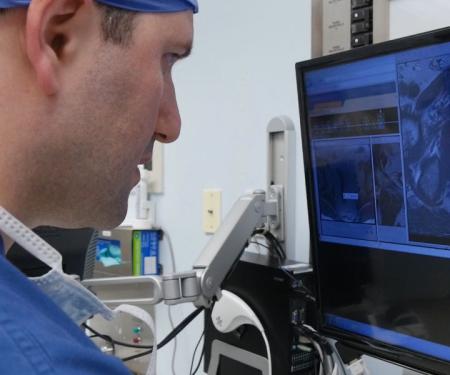
Dr. Alexander Kutikov describes the fusion biopsy system Fox Chase Cancer Center uses. It allows better assurance that tumors are located precisely in a prostate biopsy. (2:46)
At Fox Chase, we offer the latest in imaging and prostate biopsy technology in order to improve the care we can offer to our patients. Employing the power of the technique called multiparametric magnetic resonance imaging (mp-MRI), our world-class radiology team can localize tumors within the prostate with a high degree of accuracy, identifying tumors that historically went unrecognized. This technology can give patients piece of mind that aggressive disease is not being missed and uncover aggressive cancer in some individuals with tumors in uncommon and remote locations in the prostate.
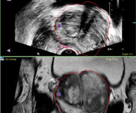
Using UroNav Ultrasound (US)/MRI fusion technology, which allows precise targeting with biopsy to evaluate lesions found on mp-MRI, we are now less likely to miss aggressive disease and, thus, potentially minimize the need for repeated prostate biopsies in the future. Furthermore, we can better assure men who choose active surveillance for low-risk prostate cancer that an aggressive tumor is not missed or overlooked. Indeed, Fox Chase is one of the first institutions in the Delaware Valley to offer US/MRI fusion capabilities to patients undergoing prostate biopsy.
If you or your loved one have an elevated PSA and are contemplating a biopsy, are due for a repeat prostate biopsy for suspected prostate cancer, or are on active surveillance for prostate cancer, we encourage you to consider getting an opinion with our prostate cancer team to see if US/MRI fusion technology can be a helpful addition to your clinical evaluation.
Why a Fusion Biopsy?
Fox Chase Cancer Center and Temple University Hospital were the first sites in the area to offer targeted MR/ultrasound fusion biopsy, a powerful diagnostic tool for the analysis, planning and targeted biopsy of the prostate. This innovative technology may result in fewer needle biopsies and better treatment decisions for patients with prostate cancer. It may also effectively detect tumors in men with prior negative biopsies but persistently elevated prostate-specific antigen (PSA) levels.
The technology fuses pre-biopsy MR images of the prostate with ultrasound-guided biopsy images in real time to create a detailed, three-dimensional view of the prostate, reducing chances that an aggressive form of prostate cancer is missed. Biopsies are mapped, targeted and tracked in a 20-minute procedure done in an outpatient setting under local anesthesia. The result is excellent delineation of the prostate, as well as any suspicious lesions.
Fusion Biopsies vs. Traditional Biopsies
In a traditional core needle biopsy, urologists use a fine needle to collect a number of tissue samples from the prostate gland, which are then examined for cell abnormalities that are a sign of prostate cancer. Whereas the traditional method lacks precision and uses random prostate sampling, MR/ultrasound fusion biopsy helps urologists to avoid missing hard-to-find, yet often aggressive prostate cancer. The technology offers the potential to minimize the over diagnosis of non-aggressive cancers, while maximizing the likelihood of detecting aggressive tumors.
Biopsies guided by MR/ultrasound fusion enable both physicians and patients to make more informed decisions ranging from prostate cancer diagnosis to treatment options—such as active surveillance or more radical interventions.
You may be a candidate if you:
- Possess an elevated PSA level
- Have a negative prior prostate biopsy with continued elevation or rising PSA
- Are on active surveillance for prostate cancer and require rebiopsy