Just as no two patients are identical, the same is true for lung cancer diagnosis. This condition can vary by type, stage, and many other factors. So an accurate diagnosis is critical for developing the best treatment plan for you.
At Fox Chase, physicians blend the most current technology with decades of experience to diagnose lung cancer of all types, from tumors to metastatic disease.
We use a variety of tests, including imaging technology and biopsy procedures, to detect and diagnose lung cancer and create a personalized plan for your treatment. These procedures are also used to find out if cancer cells have spread within the chest or to other parts of the body, a process called staging.
How is Lung Cancer Diagnosed?
An accurate diagnosis of your cancer — its type and stage — will help your physicians develop a treatment plan for you that is designed to increase your chances of recovery. We draw on the expertise of a multidisciplinary team to develop an accurate diagnosis so that we can create a treatment approach that offers the best chance of controlling your cancer, preserving lung function, and reducing the risk of recurrence.
Preliminary testing
At the beginning of the process and throughout your treatment, your doctor will do the following to diagnose the disease and assess your condition:
Physical exam and history
An exam of the body to check general signs of health, including searching for signs of disease, such as lumps or anything else that seems unusual. The physician will also take a history of health habits, including smoking, and past jobs, illnesses, and treatments.
Laboratory tests
Medical procedures that test samples of tissue, blood, urine, or other substances in the body. These tests help to diagnose the disease, plan and check treatment, and/or monitor the disease over time.
Pulmonary function test (PFT)
This test helps determine how well the lungs are working, measuring how much air the lungs can hold and how quickly air moves into and out of the lungs. The test also measures how much oxygen your body uses and how much carbon dioxide is given off during breathing. It is also called a lung function test.
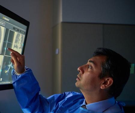
Imaging
To detect lung cancer, the following image tests may be used:
Chest X-ray
This is an X-ray of the organs and bones inside the chest. An X-ray is a type of energy beam that can go through the body and onto film, making a picture of areas inside the body. It can help physicians spot abnormalities or growths.
Computerized Tomography (CT) scan or Computerized Axial Tomography (CAT) scan
In a CT scan, special X-rays take a series of pictures of the inside of your body. Each picture displays a thin “slice” of an organ, bone, or other tissue. The entire series is put together to create 2-dimensional and 3-dimensional images, giving your physician a better idea of the location and extent of a lung tumor. A dye may be injected into your vein or swallowed to help the organs or tissues show up more clearly.
MRI (magnetic resonance imaging)
This machine uses a magnet, radio waves, and a computer to make a series of detailed pictures of areas inside the body, such as the brain. This procedure is also called nuclear magnetic resonance imaging (NMRI).
PET scan (positron emission tomography scan)
This imaging procedure can detect malignant tumor cells. The patient lies on a table that slides through a PET machine and a very small amount of radioactive glucose (sugar) is injected into the vein. The PET scanner rotates around the body and makes a picture of where glucose is being used in the body. Malignant tumor cells show up more brightly in the picture because they are more active and take up more sugar than normal cells. A PET scan and CT scan may be done at the same time (a PET-CT scan).
Radionuclide bone scan
This is a procedure to check if there are rapidly dividing cells, such as cancer cells, in the bone. In this procedure, a very small amount of radioactive material is injected into a vein and travels through the bloodstream and into the bones, where it concentrates in areas where cancer cells are present. The radionuclides give off gamma rays (similar to X-rays), which are detected by a gamma ray-sensitive camera.
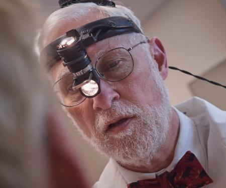
Biopsy
To determine if cancer is present, doctors often need to take tissue samples in a process called biopsy. Some techniques for getting samples include:
Fine-needle aspiration (FNA) biopsy of the lung
This involves removing tissue or fluid from the lung using a thin needle. A CT scan, ultrasound, or other imaging procedure is used to locate abnormal tissue or fluid in the lung. A small incision may be made in the skin where the biopsy needle is inserted into the abnormal tissue or fluid. A sample is removed and sent to the laboratory. A pathologist then views the sample under a microscope to look for cancer cells. A chest X-ray is done after the procedure to make sure no air is leaking from the lung into the chest.
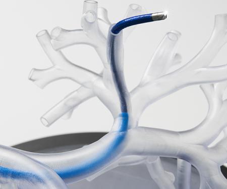
Bronchoscopy
A procedure to look inside the trachea and the two large airways (each called a bronchus; the plural is bronchi) in the lung for abnormal areas. A bronchoscope is inserted through the nose or mouth into the trachea and lungs. A bronchoscope is a thin, tube-like instrument with a light and a lens for viewing the inside of the body on video. It also has tools to remove tissue samples, which are checked under a microscope for signs of cancer.
Endobronchial ultrasound (EBUS)
A minimally invasive imaging test that uses an endoscope—a thin, tube-like instrument with a light and a lens for viewing—with a probe at the end. While the patient is under anesthesia, the endoscope is inserted into the mouth and then down into the airways. It bounces high-energy sound waves (ultrasound) off internal tissues or organs and makes echoes, which form a picture of body tissues called a sonogram. EBUS may be used to guide fine needle aspiration (FNA) biopsy of the lung, lymph nodes, or other areas.
Video-assisted thoracoscopic surgery (VATS)
A surgical procedure to look at the organs inside the chest to check for abnormal areas. An incision is made between two ribs, and a thoracoscope is inserted into the chest. A thoracoscope is a thin, tube-like instrument with a light and a lens for viewing the inside of the body on video. Like a bronchoscope, it also has tools to remove tissue samples, which are checked under a microscope for signs of cancer. In some cases, this procedure is used to remove part of the esophagus or lung (a lung wedge resection). If certain tissues, organs, or lymph nodes can’t be reached, a thoracotomy may be done. In this procedure, a larger incision is made between the ribs and the chest is opened.
Thoracentesis
The removal of fluid from the space between the lining of the chest and the lung, using a needle. A pathologist views the fluid under a microscope to look for cancer cells.
Mediastinoscopy
A surgical procedure to look at the organs, tissues, and lymph nodes between the lungs for abnormal areas. An incision is made at the top of the breastbone and a mediastinoscope is inserted into the chest. A mediastinoscope is a thin, tube-like instrument with a light and a lens for viewing. It may also have a tool to remove tissue or lymph node samples, which are checked under a microscope for signs of cancer.
Pathology
When samples have been taken, they may be subjected to the following tests:
Sputum cytology
A pathologist checks a sample your sputum, which is mucus coughed up from the lungs, under a microscope for cancer cells.
Light and electron microscopy
A laboratory test in which cells in a sample of tissue are viewed under regular and high-powered microscopes to look for certain changes in the cells that could mean cancer.
Immunohistochemistry
A test that uses antibodies to check for certain antigens in a sample of tissue. The antibody is usually linked to a radioactive substance or a dye that causes the tissue to light up under a microscope. This type of test may be used to tell the difference between different types of cancer.
How Soon Can I Expect My Results?
It usually takes at least one to two days to receive a test report, but it can be longer. The amount of time varies for each patient. Your physician will provide your test results as soon as possible, provide a thorough explanation, and answer any questions you have. From there, he or she will recommend potential procedures and treatment options that are right for your diagnosis.
What If I Have Already Been Diagnosed At Another Hospital?
If you’ve been diagnosed with lung cancer, it feels urgent to get into care immediately. However, a second opinion could lead you to new treatment options that might be more effective for your condition. In fact, it’s common for people to seek a second opinion in a cancer diagnosis, and some insurance companies even require it.
Coming to Fox Chase also means you’ll likely have greater access to clinical trials that might be appropriate for your condition. Fox Chase has been designated as a Comprehensive Cancer Center by the National Cancer Institute (NCI), meaning it’s part of a network of research centers where hundreds of clinical trials are going on every day.
If you decide to seek a second opinion at Fox Chase, bring your medical records, imaging results, and pathology report, or have your physician forward them to us. (Your pathology report is the medical report that gives a diagnosis based on tests run on tissue samples from the cancer site.) Our nurse navigators can help you manage this.
Please note that your new pathology report — the medical report we generate based on an analysis of tissue samples from the cancer site — could potentially change your diagnosis, since lung cancer and its stage are sometimes misdiagnosed. Our specialists will review your pathology report, imaging, and medical history thoroughly as they develop their diagnosis and recommend a treatment plan.
When you come for your appointment, it also can be helpful to bring a family member or friend who can help you understand and remember the information you receive.
Your Patient Care Team At Fox Chase
In order to design an individualized care and treatment plan for each patient, we draw on the diverse experience of physicians and other clinical staff in thoracic surgery, medical oncology, radiation oncology, radiology, and pathology, who work together to diagnose your cancer and help you make informed decisions about treatment. Each member of our lung cancer program is focused on delivering excellent treatment and helping you retain lung function.
“The specialists here consult on cancer cases all the time. We don’t always agree, but that’s all right. It’s reassuring to me that I can ask other people who might have a different perspective but whose opinions are absolutely worthwhile.”— Joseph Treat, MD, medical oncologist and lung cancer specialist