Once a tumor has been identified, a biopsy is necessary to obtain a tissue sample, which is required in order to make a diagnosis and to determine the cancer staging. In a traditional core needle biopsy, a fine needle to collect a number of tissue samples, which are then examined for cell abnormalities that are a sign of cancer. Fox Chase radiologists specialize in performing image-guided biopsies. Using ultrasound, CT or MRI guidance, radiologists can sample even small lesions or lesions that are difficult to access by other means, as well as biopsies on patients with complicating medical factors. All biopsies performed at Fox Chase are minimally invasive.
Related Articles
-

-
A photograph of a medical professional feeling the sides of a patient's neck.
-
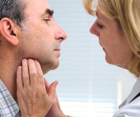
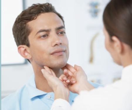
A photograph of a medical professional using her hands to feel the underside of a patient's chin.
-
-
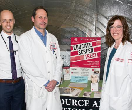
A poster reading "Oral, Head & Neck Cancer Awareness Week, free screening events hosted by the Temple Head and Neck institute.""
-
-
-
-
Oral, Head and Neck Cancer Awareness Week 2016